From Textbooks to the Tropics: How a Medical Mission Trip Transformed My Students
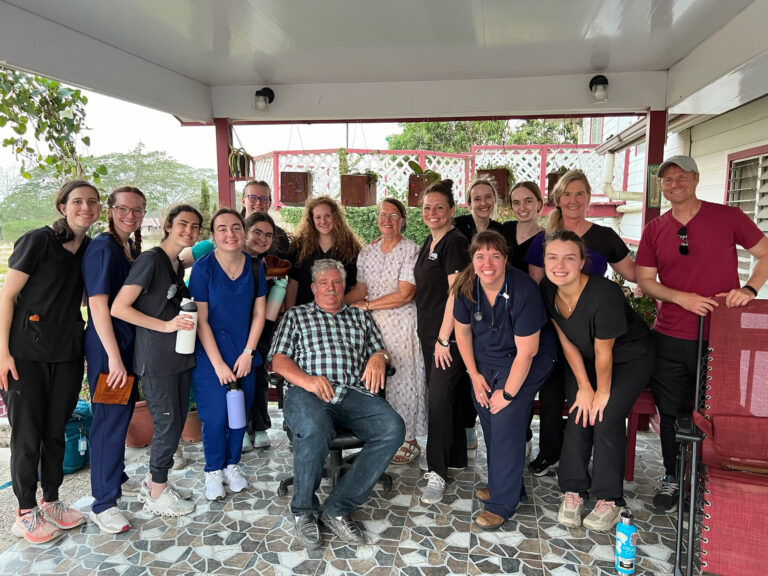
As a professor, I’ve witnessed the dedication and passion of my students daily, but sometimes, the most profound lessons happen outside the classroom. Recently, I had the privilege of co-leading a group of health science students on a medical mission…